Tendinosis
Sometimes, when an athlete’s leg boldly goes astray, the tendons connecting muscles to bones are injured in a way that does not lead to natural healing. The fibers degenerate, pain occurs, and the athlete is sidelined. Here is what’s going on.

Tendons are made up of collagen (protein) fibers interspersed with cells, vessels, nerve fibers, and water. Injuries most often occur when the tendon fibers stretch past their point of no return and break. A few fibers breaking is called a strain. Many fibers are a tear, and complete disruption is called a rupture.
When collagen fibers tear, a little or a lot, bleeding occurs. The blood and the cells of the tendon send out “red alert” signals, telling the body’s stem-cell-derived self-repair cells to race to the scene of trauma and direct the healing process. The first of these cells to arrive are called macrophages, which come in multiple forms. While M1 macrophages eat away the torn tissue ends and clean up the damage, M2 macrophages release repair-stimulating chemicals that stimulate new collagen formation. Most often this process of tendon repair unfolds over the course of 12 weeks, with further strengthening of the repair tissues over the course of a year.
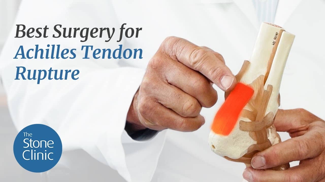
Sometimes, though, the process doesn’t work as it should. In complete ruptures, the gap between the tendon ends may be too large for the repair tissue to form a new tendon, or the tendon formed may be too long. This occurs commonly in Achilles tendon ruptures. Without surgical repair, these tendons will still heal—but with overlong fibers, leading to a weaker calf. Most surgeons repair these ruptures with the goal (especially important for the jumping athlete) of restoring the tendon’s normal length and power.
In partial tears, especially those that occur in the middle of the tendon, the blood supply and repair process can also fail, leading to an area of degenerated collagen fibers within the tendon. This is classically called tendinosis. The tendinosis fibers, curled up and without metabolic activity, wall themselves off—whereas the tendinitis fibers are inflamed and full of vessels and nerve endings. The pain of tendinosis may come from the incomplete vascular penetration and frustrated non-penetrating nerve fibers that accompany those vessels. The actual source of the pain, however, is not yet proven.
While tendinitis responds to anti-inflammatory drugs, massage, ice, and heat, tendinosis fibers languish in their degenerated state—unless a dramatic intervention occurs. In the past, injections of cortisone were tried. But as the steroid inhibits cell metabolism rather than stimulating it, those injections too commonly led to complete tendon rupture. Today tendinosis is treated by mechanical stimulation. Needling or surgical excision is used to change the biology of the locus of degeneration. This manual breaking-up of the tissue is then augmented with growth factor stimulation from platelet-rich plasma (PRP) and other growth-factor-rich sources. These substances, rich in anabolic factors that promote cell metabolism and tissue growth, also act as natural pain relievers.
In controlled studies of growth factor augmentation after mechanical tissue disruption, these solutions permitted athletes to return to sports faster than saline or exercise alone. Yet even with these modern treatments, the collagen fibers still required a full year to increase in strength. We still have a long way to go before we can wave a Star Trek-style Tricorder over an injury and induce an immediate injury resolution—but we are working on it.
What's the Best Way to Fix a Ruptured Achilles Tendon?
At The Stone Clinic, we repair Achilles tendon ruptures without open surgery using the Percutaneous Achilles Repair technique. Over the past 30 years, Kevin R. Stone, MD, has refined this approach to eliminate the need for a large incision, preserve growth factors that accelerate healing, and reduce infection risk. Here’s how.