Achilles Repairs for Top Level Athletes
Hear From Our Patients
Surfers Returns from Ruptured Achilles with Percutaneous Repair
Pop goes Achilles. Almost every victim says they heard, or felt, a shot in the back of their calf. The prospect of a ruptured Achilles strikes fear in everyone, especially professional athletes. Here is our update.
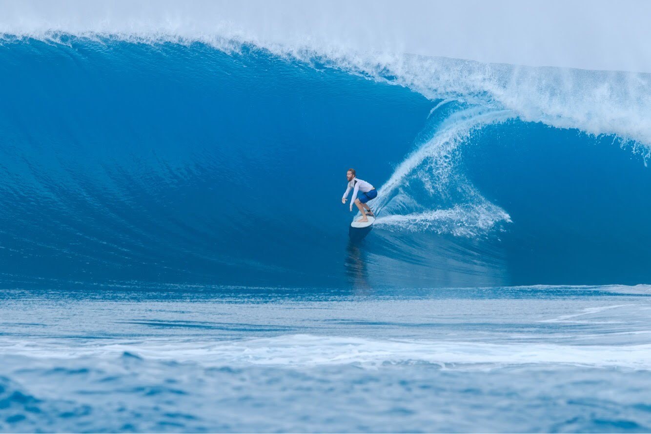
Pictured: Stone Clinic patient Harper C., surfing over 100 days last year after percutaneous Achilles repair.
The Achilles is a broad, flat tendon that travels in a sheath from the calf muscles to the calcaneus (heel bone) at the bottom of the foot. It is designed to stretch as the calf muscles contract during walking, providing power for the push-off from the toes. It has a great blood supply and nerve supply, which permits it to heal on its own without surgery.
On complete rupture, however, the ends of the tendon separate and a gap is felt when the Achilles is examined. That gap, if left open, will eventually heal with scar tissue but the tendon will be longer than it was before the injury. This increased length accounts for its reduced power. For this reason, almost all athletes select tendon repair over a “wait-and-see.”
Repair of the Achilles was commonly done as an open surgical procedure as soon as possible after injury. The incision directly over the Achilles tendon sliced through the thin sheath and released the natural hematoma (blood clot) that formed at the torn tendon ends. Sutures were then applied, sewing the tendon ends together. Casts or a brace were often worn, for a period of weeks or months, to permit the surgical repair to heal.
The problem with this technique is that the sheath contains sensitive nerves and fine blood vessels, while the newly formed hematoma—which is bathing the ends of the ruptured tissue—holds the body's natural healing stem cells. This loss of both the sheath and the newly formed blood clot delayed healing and created an unnaturally high infection rate with scar formation after open Achilles repair.
Griffen and Ma in 1977 designed a percutaneous technique for sewing the tendon ends together without opening the skin, thereby preserving the hematoma. We modified this technique in 1990, using completely resorbable sutures placed beneath the skin in a crisscrossed, reinforced fashion.
Our healing tendons were monitored with sequential MRI which, over a period of years, demonstrated that they healed relatively rapidly and with hypertrophy (increased size of the tendon itself). This permitted us to accelerate our rehabilitation programs—especially important for career athletes trying to return to sport as rapidly as possible. We still don't know how fast we can actually push the recovery, although we often learn the most from our patients who “cheat.” Patients who don't follow our advice to protect the tendon during the first couple of months, and send us pictures of themselves walking and exercising unprotected, remind us that the body can heal on its own as long as it has a little bit of help. In this case, “help” is the sutures that bring the tendon ends together (which usually dissolve completely by six weeks).
Additionally, the injection of PRP and growth factors (which we harvest from a blood draw from the patient and inject around the healing tissue during the initial window after surgery) may be responsible for further accelerating the natural healing process.
Other techniques that have been developed, using percutaneous devices and permanent sutures and anchors. We have not found any of these to be necessary; in fact, they present a risk should the patient develop an infection or other scarring problems.
The Achilles remains a devastating injury for athletes during their season. However, it can be repaired without open surgery and rehabilitated relatively rapidly when augmented by growth and recruitment factors that bring the body's own stem cells to the site of healing. The ruptured Achilles patient can look forward to a full career on the repaired tendon. We only wish that Achilles’ mother had held him by the great toe.
Dr. Stone Explains the Percutaneous Surgical Technique
At The Stone Clinic, we repair Achilles tendon ruptures without open surgery using the Percutaneous Achilles Repair technique. Over the past 30 years, Kevin R. Stone, MD, has refined this approach to eliminate the need for a large incision, preserve growth factors that accelerate healing, and reduce infection risk. Here’s how.