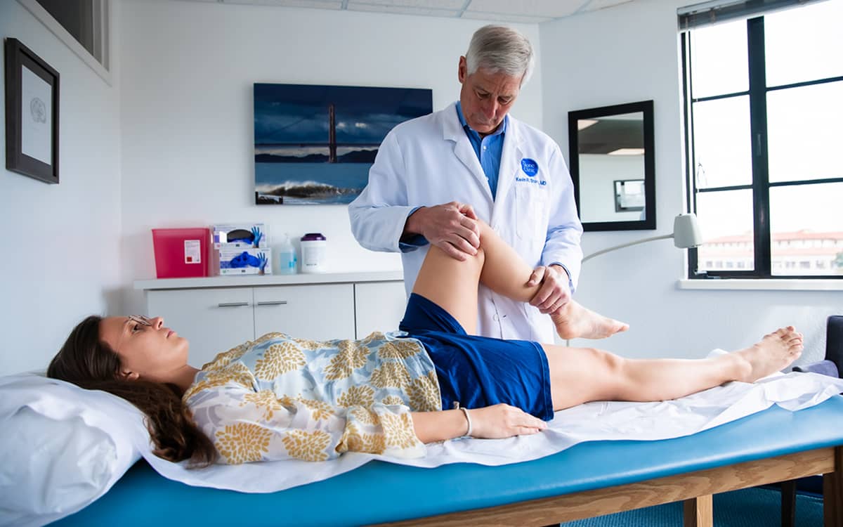
Why I Need To Physically Examine Patients
“Doc, can’t you just look at my knee X-ray and tell me what procedure I need?” Yes, I sometimes can, but that is not the best way. Here is why.

People vary. Their knee joints are remarkably unique to their body, to their history of use, to their previous injuries, to the ligaments that guide their motion, and to the meniscus and articular cartilage that absorb their body weight while they are walking and running. Understanding the nuances of each person’s knee permits me to guide them and operate on them as individuals, customizing each step for their best outcome.
The first step in my assessment is observing their gait. Shortly after our initial introduction, I watch them walk (and sometimes run) up and down the hallway near my office. How people walk—how their hips, knees, ankles, and feet all work together—tells much of their story. The bowlegged person who is only bowed on one side differs greatly from the person born with both bowlegs. The person who limps only when they run (and may not even be aware of it) has muscle engagement quite different from those who don’t limp. People with ankles that don’t bend and hips that don’t rotate might say they have knee pain—but the main problem may lie elsewhere. Woe be the surgeon who operates on the knee when it was the arthritic hip referring pain to the knee joint.
The patient’s history can now be obtained remotely, and augmented by our AI voice agent listening in, but the physical exam itself must be done in person. How the knee bends, how it rotates, how stable it is in multiple planes—both in full extension and in various degrees of bending—can be assessed only by experienced hands feeling the changes that occur during the exam. Reproducing the pain by applying finger pressure or by angling the knee during the range of motion, tells me much about which specific tissues are catching, loose, or torn, thus generating pain.
The next step is imaging. Here, the combination of an X-ray and MRI for assessment of the knee joint (and for most joints) is the gold standard. While an MRI adds vast amounts of soft tissue information, the X-ray provides bone and joint space measurements more accurately. This is because while the MRI is obtained sitting (without putting a load on the joint) the X-ray is taken while standing. (Ultrasound is sometimes also useful, but nowhere near as helpful for most knee joint diagnoses.) Still, determining where the knee grinds and where it is swollen is not revealed by the X-ray alone.
Next the rehabilitation team steps in, guided by a program that is an extension of my eyes, ears, and hands. They assess the patient’s understanding of the advice they have been given and push the patient through a series of exercises that help them understand where they are strong, where they are weak, and how to uncover other physical attributes that can guide both their treatment and their rehabilitation program. Our goal, as always, is to help them return to their lives and activities not just repaired, but better than they have been in years.
So if you want my best advice, here it is: Let me use all of my tools to assess your knees, shoulders, ankles, sports injuries, and arthritis. I love giving advice based on solid data, rather than on information conveyed through the virtual ether.
